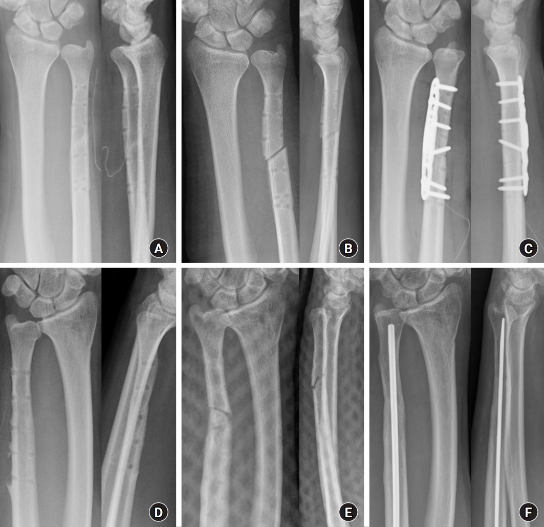
Comparison of ulnar shortening osteotomy for idiopathic ulnar impaction syndrome using conventional or ulnar osteotomy plates and with or without interfragmentary screw fixation
Article information

Abstract
Purpose
This study investigated the impact of plate type on the clinical and radiological outcomes of ulnar shortening osteotomy (USO) by comparing conventional and ulnar osteotomy plates. The effect of interfragmentary screw fixation (ISF) during USO was also assessed.
Methods
Seventy-eight patients were divided into three groups according to the type of plate: 3.5-mm dynamic compression plate (DCP), 3.5-mm limited contact DCP, and 2.7-mm locking compression plate ulna osteotomy system (all from Depuy-Synthes). The patients were also divided into two groups according to whether ISF was performed. Clinical and radiological outcomes, including time to bone union, presence of delayed union, and refracture after hardware removal, were analyzed. Other factors that might affect bone union, such as smoking and underlying diseases, were also evaluated.
Results
No significant differences were found in clinical and radiological outcomes according to the type of plate. Eight of 51 patients (15.7%) in the without-ISF group showed delayed bone union. Forty-three patients in the without-ISF group underwent hardware removal, and refracture due to low-energy trauma after hardware removal was observed in five of those 43 patients (11.6%). Bone union time was significantly shorter in the with-ISF group (7.6±2.7 weeks vs. 9.8±6.6 weeks). Diabetes mellitus and ISF were associated with the delayed bone union.
Conclusion
The plate type had no influence on the clinical and radiological outcomes of USO in patients with idiopathic ulnar impaction syndrome. However, ISF during USO has several advantages, such as early bony union and prevention of refracture after hardware removal.
Introduction
Ulnar impaction syndrome (UIS) can cause medial wrist pain related to excessive weight bearing across the ulnar aspect of the wrist. It occurs when the ulnar head abuts the triangular fibrocartilage complex and carpus, resulting in ulnar-sided wrist pain [1]. Moreover, idiopathic UIS is generally seen in patients with static or dynamic positive ulnar variance with wrist pronation and forceful grip [1,2]. Ulnar shortening osteotomy (USO) was first described as an operative intervention to decompress the ulnocarpal articulation by Milch [3] in 1941. Since then, it has been widely used in clinical practice and various modified surgical methods have been introduced. Although USO has been successfully used for the treatment of idiopathic UIS [4-6], the surgery carries the risk of hardware irritation, delayed union, and nonunion [7,8].
The purpose of this study was to compare the clinical and radiological outcomes of conventional and ulnar osteotomy plates for USO in patients with idiopathic UIS. In addition, the effect of interfragmentary screw fixation (ISF) in USO was assessed.
Methods
Ethics statement: This study design was approved by the Institutional Review Board of Dankook University Hospital (No. 2022-09-013). The study was performed in accordance with the Declaration of Helsinki, and written informed consent was obtained for publication of this article and accompanying images from the patients.
From March 2008 to May 2016, 78 patients who underwent USO using plate fixation were retrospectively reviewed. Among the patients who were diagnosed with UIS and took the USO, anyone who was diagnosed with secondary UIS and had less than 1 year of the follow-up period was excluded. All patients were evaluated and treated by a single specialized hand surgeon. Three types of plates were consecutively used: 3.5-mm dynamic compression plate (DCP; Depuy-Synthes, West Chester, PA, USA), group I (n=31); 3.5-mm limited contact DCP (LC-DCP, Depuy-Synthes), group II (n=19); and 2.7-mm locking compression plate (LCP) ulna osteotomy system (Depuy-Synthes), group III (n=28). And also, patients were divided into two groups based on whether they underwent ISF: with-ISF group (n=27) and without-ISF group (n=51).
Clinical outcomes were respectively evaluated by DASH (Disability of Arm, Shoulder and Hand) and PRWE (Patient Related Wrist Evaluation) scores preoperatively and 1 year after surgery. In addition, radiological outcomes, including time to bone union, presence of delayed union, and refracture after hardware removal were assessed. Bone union was defined radiologically when callus formation was observed on three of the four cortices on anteroposterior and lateral plain radiographs. Furthermore, the delayed union was defined when the union was not observed until 12 weeks. Other possible factors that might affect bone union, such as smoking and underlying diseases (that is, diabetes mellitus) were also assessed.
1. Surgical procedures
The USO was performed by a single specialized hand surgeon. Before performing USO, the author planned the necessary amount of resection to restore the ulnar variance between 0 and –1 cm. On the distal ulnar-sided forearm, 7 to 8 cm of skin incision was placed. The ulna was approached by dissecting between the flexor carpi ulnaris and extensor carpi ulnaris, while preserving the dorsal sensory branch of the ulnar nerve. After that, plate was temporarily applied using guide pins to predrill the two distal holes and one proximal hole. After removing the plate, an oblique-cut osteotomy was performed considering the edges and rotation without any cutting guide or additional apparatus. The plate was placed using a reduction clamp to achieve anatomical reduction, and then screws were firmly secured. All patients who had undergone USO were applied protective long arm splint for 2 weeks, short arm cast for 4 weeks, and wrist brace for 6 weeks, postoperatively.
2. Statistical analysis
Categorical variables were compared using the chi-square test and continuous variables were compared using the independent t-test and one-way ANOVA. Factors potentially associated with bone union time were analyzed using a logistic regression analysis. Statistical analyses were performed using IBM SPSS Statistics (IBM Corp., Armonk, NY, USA). Results with p-values that were less than 0.05 were considered for statistical significance.
Results
The average follow-up time was 15.8 months (range, 12–27 months). There were 43 male and 35 female patients with a mean age of 42 years (range, 22–69 years). Out of 78 patients, there were no significant differences in clinical and radiological outcomes according to the types of plates (Table 1). When comparing groups according to whether ISF was performed, there was no significant difference in demographic factors and clinical outcomes between the two groups. However, bone union time was significantly shortened from 9.8±6.6 to 7.6±2.7 weeks in the with-ISF group. Delayed bone union was confirmed only in the without-ISF group. Eight of the 51 patients (15.7%) who were treated without interfragmentary fixation showed delayed bone union. Unlike the with-ISF group, refracture by low-energy trauma after hardware removal was observed in five of 43 patients (11.6%) in the without-ISF group. However, this difference was not significant (Table 2). Moreover, to identify the risk factor related to delayed bone union, factors potentially associated with bone union time, such as sex, smoking, diabetes mellitus, ISF, and the type of plates were analyzed using logistic regression analyses. Among the risk factors which are related to bone union, diabetes mellitus (odds ratio [OR], 4.86; p=0.046) and ISF (OR, 2.44; p=0.042) were associated with the delayed bone union.
Clinical and radiological outcomes according to the type of plate

Clinical and radiological outcomes according to interfragmentary screw fixation
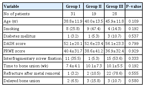
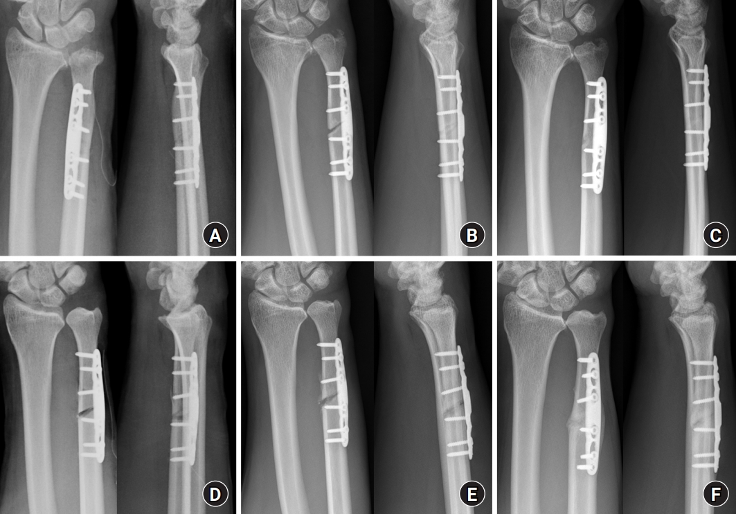
All patients showed bone union at the final follow-up. Eight patients who showed delayed bone union were treated conservatively; thereafter, they showed radiological union at a mean period of 22.2 weeks (Fig. 1). Five patients who showed refracture after hardware removal underwent revision open reduction and internal fixation (n=4) or closed reduction and intramedullary nail fixation (n=1), and bone union was achieved at the final follow-up (Fig. 2).
(A) A 62-year-old man was treated with a 2.7-mm locking compression plate (LCP) ulna osteotomy system (Depuy-Synthes, West Chester, PA, USA), and interfragmentary screw fixation was not performed. Immediately postoperatively, a neutral ulnar variance was achieved and the ulnar osteotomy plate settled well. (B) Twelve weeks later, the gap at the osteotomy site had increased, and delayed bone union occurred. (C) Twenty-four weeks later, radiological bone union was spontaneously achieved. (D) A 59-year-old man was treated with a 2.7-mm LCP ulna osteotomy system (Depuy-Synthes), and interfragmentary screw fixation was not performed. (E) Thirteen weeks later, callus formation was seen around the osteotomy site. (F) Twenty-six weeks later, radiological bone union was achieved with excessive callus formation.
(A) A 49-year-old woman underwent ulnar shortening osteotomy without interfragmentary screw fixation. Eleven months later, solid bone union was achieved and hardware removal was done. (B) Refracture occurred by low-energy trauma after hardware removal. (C) A revisional operation was done with ulnar osteotomy plate and interfragmentary screw fixation was performed. Seven weeks later, solid bone union was achieved. (D) A 29-year-old man underwent ulnar shortening osteotomy without interfragmentary screw fixation. Twelve months later, solid bone union was achieved and hardware removal was performed. (E) After hardware removal, refracture by low-energy trauma occurred. (F) Revisional closed reduction and intramedullary nail fixation were performed. Nine weeks later, solid bone union was achieved.
Discussion
As a result of positive ulnar variance, overloading between the ulnar carpus and the distal ulna occurs. Hence, patients usually report ulnar-side wrist pain, diminished grip strength, swelling, and limitation of wrist motion. USO is the preferred treatment option for correcting positive ulnar variance, which relieves pain by restoring neutral or slightly negative ulnar variance. Jungwirth-Weinberger et al. [9] reported that using 2.7-mm LCP ulnar osteotomy system plate for USO in patients with UIS displayed shorter bone healing time than that resulting from the use of 3.5-mm LC-DCP. However, in our study, there were no significant differences in either clinical or radiological outcomes according to the types of plate.
Hulsizer et al. [10] reported that 12 out of 13 patients achieved complete pain relief at a mean of 2.3 years with freehand transverse osteotomies and four out of 13 had postoperative complications. On the other hand, Chun and Palmer described a series of 30 cases of an oblique freehand osteotomy technique with good results, although there were few complications, including no nonunion [5]. Theoretically, when the interfragmentary screw crosses the fracture line, the threads engage the fragment and the lag effect of the screw reduces the fracture gap. Further tightening of the lag screw pulls the distal fragment in a proximal direction, compressing the fracture site and enhancing fracture stability, which results in improved fracture healing [11]. Hence, in this study, USO was performed using the oblique osteotomy technique in all patients. Previous studies reported 0% to 18% of nonunion [12-16]. However, the incidence of the nonunion and delayed union in this present study was 0% and 10.2%, respectively, which are lower rates than most reported in the literature. A study by Chen et al. [17] demonstrated that 30% of smokers compared to 0% of nonsmokers developed nonunions. Moreover, Gaspar et al. [18] revealed that smoking and diabetes mellitus were significantly elevated risks for both delayed union and nonunion in USO. However, in this present study, only diabetes mellitus and ISF were significantly associated with the delayed bone union.
Pomerance [19] insisted that local pain or irritation was the main cause of plate removal, and demonstrated a 2.5% incidence of refracture after hardware removal (mean time to hardware removal, 6.6 months) after USO. Furthermore, according to Mih et al. [20], 11.3% of patients experienced refracture after hardware removal (mean time to hardware removal, 19 months) from various forearm pathologies. We demonstrated a 78.2% (61 of 78) incidence of hardware removal in this study, which is comparable to the results of other studies. Among them, only five patients (8.2%) experienced a refracture after hardware removal. All patients underwent revision surgery, and the bone union was achieved at the final follow-up.
This study had several limitations. First, the retrospective nature of this study could have introduced selection bias. The author suggests that a randomized comparative study would help elucidate the most important factors. Second, the degree of diabetes mellitus control has not been investigated in detail. These details may have been partly responsible for the differences in the results. Third, in this study, not like the category of delayed bone union, refracture after metal removal showed no significant difference between the with-ISF group and the without-ISF group. As refracture after metal removal could be considered a serious complication, this could undermine claims about the effectiveness of ISF. However, unlike the without-ISF group, which showed five cases of refracture (11.6%) after metal removal, none (0%) occurred in the with-ISF group. Hence, although a significant difference was not achieved, it was assumed that it could sufficiently support the effectiveness of ISF.
Conclusion
Plate types did not affect the clinical and radiological outcomes of USO in patients with idiopathic UIS. However, ISF during USO has several advantages, such as early bony union and prevention of refracture after hardware removal. In conclusion, ISF during USO can be a good surgical option for patients with UIS.
Notes
The authors have nothing to disclose.
Funding
None.