Surgical treatment of multiple plexiform schwannomas arising from the superficial radial nerve: a case report
Article information

Abstract
Schwannoma, or neurilemmoma, is a benign neoplasm that arises from Schwann cells, which surround peripheral, cranial, and autonomic nerve sheaths. Schwannoma has been reported to occur mainly as a singular lesion of the sacral nerve or sciatic nerve in young adults. Plexiform schwannoma, a subtype of schwannoma, is a rare neoplasm known to account for 2% to 5% of total schwannomas. Schwannoma of the upper extremities is relatively rare and is reported to occur mostly in the ulnar nerve. We report, with a literature review, a case of 4.2-cm and 2.8-cm symptomatic multiple plexiform schwannomas that occurred in the superficial radial nerve and were treated without neurologic sequelae by surgical resection.
Introduction
Schwannoma, or neurilemmoma, is a benign neoplasm that arises from Schwann cells that surround the peripheral, cranial, or autonomic nerve sheaths. Schwannoma has been reported to occur mainly as a singular lesion of the sacral nerve and sciatic nerve of a young adult [1]. Plexiform schwannoma, a subtype of schwannoma, is a rare neoplasm known to occupy 2% to 5% of total schwannomas. It is most prevalent in adults in their 2nd to 5th decades of life. Most are well demarcated and are sized less than 2 cm, showing asymptomatic, slow growth [1,2]. However, plexiform schwannomas resulting from a single nerve with a greater than 2 cm and accompanied by symptoms are extremely rare [3]. Authors experienced and treated successfully a case of multiple plexiform schwannomas which is symptomatic and 4.2 cm, 2.8 cm sized in the superficial radial nerve.
Case report
The patient was a 66-year-old female. Since 10 years ago, masses were present in her left elbow joint and her forearm. No assessment or treatments were conducted. She presented to our clinic with two enlargement of the masses and tingling sensations around the masses. Her symptoms were minimal, and the masses were assessed as benign neoplasm. As a result, she was asked to follow up in 6 months of careful observation. However, worsening of the symptoms and enlargement of the masses led her to return to the outpatient clinic (Fig. 1).
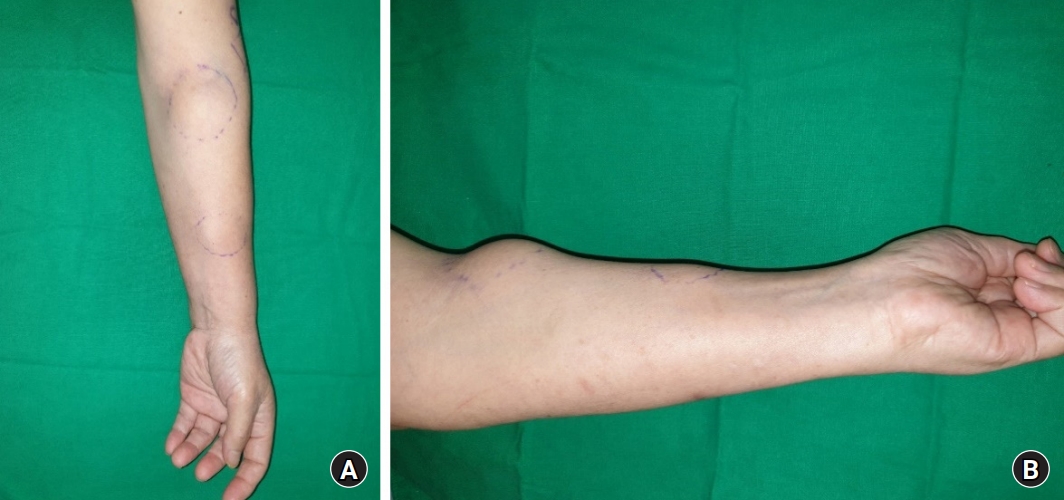
Preoperative clinical photographs of the patient. Multiple masses presented in the anterolateral aspect of patients left forearm. (A) In the anterior view, each sized 5 cm and 3.5 cm, circular masses were located in the anterior side of proximal and distal area of forearm. (B) In the lateral view, large size mass was located in the proximal area of forearm.
Initial examination reported pain of visual analog pain scale 3, tenderness and tingling sensations around the masses. Tinel’s sign was present around the dorsal aspect of the first web space of affected hand, where the superficial radial nerve distributes. Motor power of the elbow joint, carpal joint, and hand was normal. No significant findings were observed on the simple radiographs. An ultrasound reported two neurogenic neoplasms, each sized 4.2 cm and 2.8 cm and located in the anterior side of the left elbow joint and the left forearm (Fig. 2). Magnetic resonance imaging (MRI) study reported two neurogenic neoplasms, each sized 4.5×3.1 cm and 2.8×2.1 cm, located in the anterior side of the left elbow joint and the left forearm. Both masses had originated from the superficial radial nerve (Fig. 3).
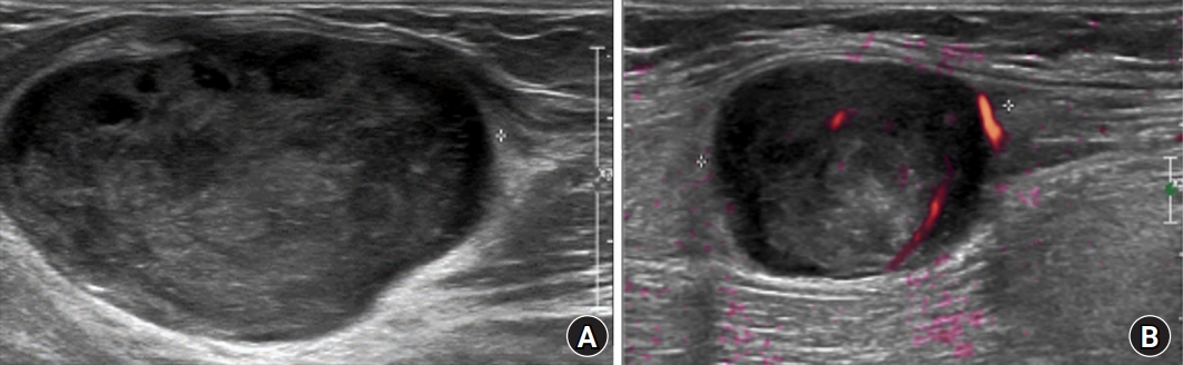
Ultrasound images. (A) An ovoid, hypoechoic lesion with a length of 4.2 cm was observed in the anterior aspect of the left elbow, connected to the superficial radial nerve on an ultrasound examination. (B) An ovoid, hypoechoic lesion with a length of 2.8 cm was observed in the anterior aspect of the left forearm, located near the median nerve, but the connection was uncertain based on the ultrasound examination.

Magnetic resonance images at the level of the forearm demonstrate areas of low signal intensity on T1-weighted imaging and intermediate to high signal intensity on T2-weighted imaging that are connected to the superficial radial nerve. (A) A mass with a length of 4.5 cm is observed on the anterior aspect of the elbow. (B) A mass with a length of 3.0 cm is observed on the anterior aspect of the forearm. (C) Two masses are observed on the sagittal view.
The operation was conducted under a nerve block of the axillary upper arm. After a linear skin incision was made over the mass site, the superficial radial nerve including the mass was exposed, and then the epineurium enclosing the mass was incised longitudinally to exfoliate the mass easily. Then, the mass was removed and the epineurium was sutured with 6-0 Prolene (Ethicon, Somerville, NJ, USA) suture material (Fig. 4). The same procedure was done on the distal mass. Pathologic examinations following a hematoxylin and eosin stain reported the Antoni A area with regular patterns and cellularity and the Antoni B area with low cellularity (Fig. 5). A well-defined multinodular bipolar mass was verified under a Masson’s trichrome stain, and a strong positive was verified under immunohistochemical staining (Figs. 6, 7). Both masses were diagnosed as plexiform schwannoma.
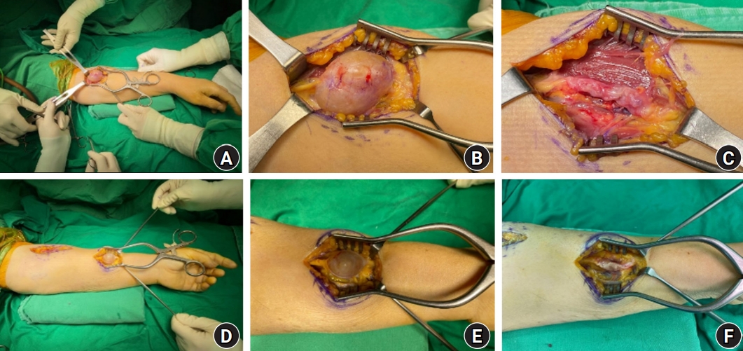
Intraoperative clinical photographs of multiple schwannomas of the upper arm at the level of the elbow (A–C) and the anterior aspect of the mid-forearm (D–F).
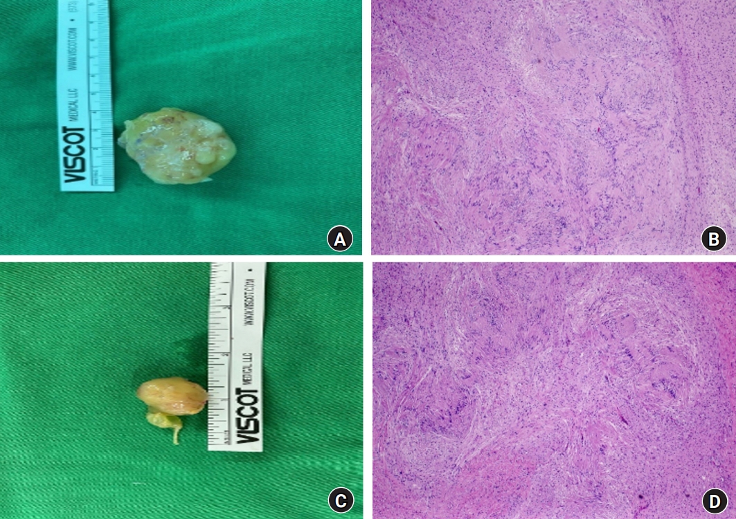
Light microscopic findings and gross mass images of the left elbow (A, B) and forearm (C, D). The tumors are cases of plexiform neurilemmomatosis, showing multilobulated nodules with hypercellular Antoni A areas and hypocellular Antoni B areas (hematoxylin and eosin stain, ×40).
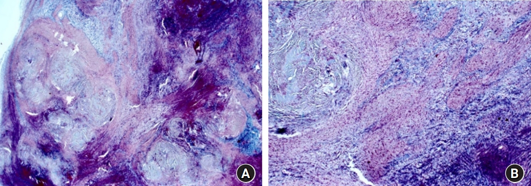
Well-defined multinodular and biphasic tumor (Masson’s trichrome). (A) ×15, (B) ×40.

Immunohistochemical staining of tumor nodules shows strong positivity for S-100 protein (×40). (A) Mass on the anterior aspect of the elbow. (B) Mass on the anterior aspect of the forearm.
A splint was applied after surgery. The splint and suture materials were removed 2 weeks later after surgery. The patient recovered her full range of motion and returned to her daily activities. The Weber’s two-point discrimination test by calipers on the affected site and normal site were 9 mm and 6 mm, respectively at the postoperative 18 months. There was sensory impairment slightly, but near normal category.
Written informed consent for publication of the clinical images was obtained from the patient.
Discussion
Schwannoma is a benign soft tissue neoplasm, arising from the nerve sheath cells (Schwann cells) of the nerve sheath. It is known to mainly occur in adults in their 3rd to 6th decade of life [4]. Sex does not seem to affect the prevalence of schwannoma. As a benign neoplasm that arises from the peripheral nerves, schwannoma requires differentiation from neurofibroma. Unlike the latter, the former is surrounded by a sheath. Notably, plexiform schwannoma, a subtype of schwannoma and a rare type of neoplasm known to constitute 2%–5% of total schwannoma, was named after the plexiform nerve sheath cells between the dermis and the subcutaneous tissues [2]. Plexiform schwannoma is prevalent in adults in their 2nd to 5th decade of life, and most show asymptomatic and slow growth [5]. While reports suggest that schwannoma can originate from any nerve, it is known to most often originate as a single lesion in the portion where flexion occurs at the extremities, especially the sacral nerve and the spinal nerve [6]. Twelve to nineteen percent of the total schwannoma occurs in the upper arms, among which mostly originate from the ulnar nerve and the median nerve [3]. Most plexiform schwannomas are asymptomatic and less than 2 cm in size. And, symptomatic plexiform schwannomas with a large size of more than 2 cm have been reported very rarely. Symptomatic large-sized multiple plexiform schwannomas that occur simultaneously at least two masses in a single nerve are extremely rare [1,6]. However, in this case, the size was 4.2 cm and 2.8 cm, which is much larger than typical tumor size originating from single nerve and is symptomatic with pain, tenderness, sensory change, and Tinel’s sign.
Ultrasound images and MRI study is reported to help diagnosis of schwannoma. Schwannoma is identified as a well-demarcated round mass in an ultrasound. The internal portion may be heterogeneous or homogenous and is connected to adjacent nerves. T1-weighted MRI is reported to show isointense signals, while T2-weighted MRI is reported to show heterogenous signal intensities with hyperintense signals [7]. Stull et al. [8] reported that signs such as nerve entering and existing sign, splint fat sign, and target sign can help the diagnosis of schwannoma. While not essential for the treatment of schwannoma, surgical resection is the only method to reach a pathologic diagnosis. Pathologically, schwannoma can be separated into areas Antoni A and B. Antoni A area is composed of spindle-shaped cells. Strong positive results for S-100 protein are obtained from the immunohistochemical staining, which is a sign specific to schwannoma as opposed to neurofibromatosis, where such results are absent. Since it is common for schwannomas to form a sheath, careful dissection from the originating nerve can lead to the excision of the neoplasm with minimal damage to the nerve. Careful surgical resection is needed since postoperative neural damage may be induced if the nerve perforated the center of the neoplasm.
Authors experienced and treated without neurologic sequelae by surgical resection a case of multiple plexiform schwannomas which are 4.2 cm and 2.8 cm sized in the superficial radial nerve have pain, tenderness, sensory impairment, and Tinel sign, and report it with literature.
Notes
The authors have nothing to disclose.
Funding
None.