Segmental schwannomatosis in the upper extremity: a case report and review of the literature
Article information

Abstract
Schwannomas, the most frequently occurring benign tumors of the peripheral nerve sheath, generally remain as painless swellings for several years before diagnosis. Multiple schwannomas involving different nerves within the same extremity are rare. We report a rare case of a 61-year-old female patient who presented with multiple schwannomas in the palmar common and proper digital nerves, 15 years after the resection of a median nerve schwannoma within the same upper extremity. Using pre-established diagnostic criteria, she was diagnosed with segmental schwannomatosis. After careful surgical resection, biopsy confirmed the diagnosis, and she recovered without neurological symptoms or limitations in the range of motion. A literature review revealed only four case series on segmental schwannomatosis, indicating its rarity. Postoperative sensory deficits are more likely in cases with multiple schwannomas in the common and proper digital nerves. We demonstrate that such complications can be avoided by meticulous dissection and separation of the tumors from the nerve fibers.
Introduction
Schwannomas (or neurilemmomas) are the most frequently occurring benign tumors of the peripheral nerve sheath [1,2]. Commonly occurring in the fourth–sixth decades of life without any sex predilection [3,4], they are mostly observed in the volar aspect of the forearm and hand. They reportedly comprise 12% of all soft-tissue neoplasms of the hand [1-3].
Because schwannomas are generally manifested as painless swellings with less common neurological deficits, the time to their diagnosis can vary from a few months to several years [4]. Magnetic resonance imaging (MRI) and ultrasonography are helpful in their diagnosis and localization [1,2]. Schwannomas are usually solitary lesions, and multiple schwannomas of different nerves within the same extremity are rare [3]. No malignant transformations of schwannomas have been reported yet; however, 8.3% of patients have experienced a local recurrence after resection [3].
We report a patient who presented with multiple schwannomas that occurred in the common and proper digital nerves on the third and fourth rays following the resection of a median nerve schwannoma in the same upper extremity 15 years ago. This patient was diagnosed with segmental schwannomatosis of the upper extremity. We have reviewed the literature and discussed this rare presentation, with hope that our findings serve as a guide to fellow surgeons and physicians who encounter similar situations.
Case report
A 61-year-old female patient presented to the outpatient clinic with tenderness and a gradually enlarging mass in the third palmar web-space of the right hand. Although the mass was first recognized 3 years ago, it was small and painless; therefore, no further examinations were performed at the time. The patient had a history of a single large schwannoma (8.0×3.5×2.6 cm) in the ipsilateral median nerve at the level of the distal forearm (Fig. 1), which was diagnosed by biopsy following its surgical resection 15 years ago. The previous surgical site had no signs of recurrence.
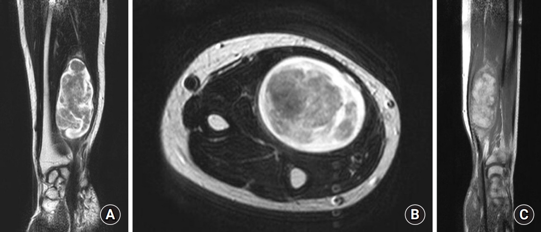
Magnetic resonance imaging scans (taken 15 years ago) reveal an 8×5-cm schwannoma encapsulating the median nerve in the right wrist. T2-weighted coronal (A) and axial (B) images show bright enhancement mainly in the tumor’s edge, while the remaining region appears to emit an amorphous low signal. On T2-weighted gadolinium enhancement (C), the low-signal areas show inhomogeneous enhancement.
At the current presentation, X-ray examination revealed no specific lesions. Although MRI detected multiple peripheral nerve sheath tumors of the digital nerve, it was necessary to discriminate against multiple tenosynovial giant cell tumors (Fig. 2). A resection biopsy was planned and a V-shaped incision was made in the third web-space to access the lesion, following which multiple tumors in the subcutaneous layer were identified easily. With the fingers slightly flexed in order to prevent stretching of the digital nerve, we performed the dissection under loupe magnification. To avoid nerve damage due to the presence of multiple lesions, we attempted to differentiate between the proximal and distal nerve fibers and the tumors and also determined the relationship between the normal nerve fibers and the tumors. The epineurium was longitudinally incised in the direction of the mass opposite to the nerve fiber, and the masses were peeled off and enucleated. Thereafter, we confirmed that the common digital nerve and the third and fourth proper digital nerves were not damaged and retained continuity (Fig. 3). A total of four tumors were noted: two were attached to the common digital nerve (tumor sizes, 2×3 cm and 1×1 cm), while one was present in the third and fourth proper digital nerves each (tumor size, 0.5×0.5 cm for both).

Preoperative magnetic resonance imaging reveals multiple masses encapsulated by the common and palmar digital nerves in the palmar aspect of the right hand. A T2-weighted short tau inversion recovery coronal image (A) reveals heterogeneous signal intensities for multiple masses. The T1-weighted axial images (B and C) reveal masses with low-signal intensities along with the digital nerve routes.
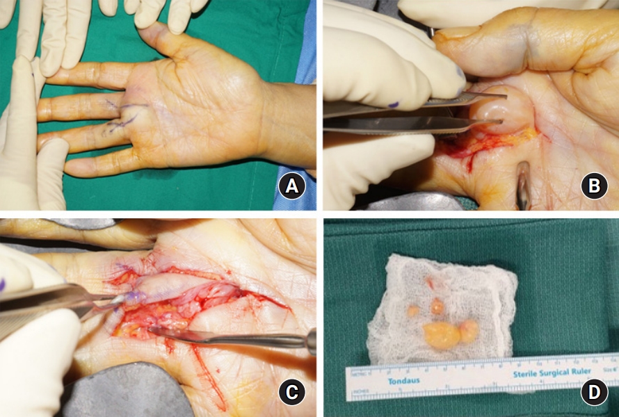
Intraoperative image taken during excisional biopsy of the mass. (A) A V-shaped incision is made along with the mass. (B) The mass is observed to be encapsulated by the palmar common digital nerves. (C) After careful excision, the nerve fiber maintains continuity. (D) A total of four masses are excised.
Gradual range of motion was allowed after applying a short arm splint for 1 week postoperatively. No neurological symptoms were observed after the operation and recovery progressed without any significant complications. On postoperative biopsy, multiple schwannomas were noted (Fig. 4). These schwannomas recurred in the ipsilateral upper limb, although not at the same site as the one located 15 years ago, and satisfied the diagnostic criteria for segmental schwannomatosis. There were no complications, including recurrence, neurological deficits, and a limited range of motion at the one-year follow-up (Fig. 5).

A low-power microscopic view of multiple resected tumors shows a multinodular growth pattern consistent with plexiform schwannomas (H&E, ×40) (A). (B) A high-power microscopic view shows spindle-shaped tumor cells forming a Verocay body made of typical nuclear palisading regions and an anuclear zone (H&E, ×200).

(A, B) Postoperative images reveal neither a limited range of motion nor any neurological deficits.
The patient provided informed consent for reporting her case with accompanying images.
Discussion
Our patient presented with multiple schwannomas in the common and proper digital nerves of the palm 15 years after a solitary schwannoma was excised from the distal forearm. Although the previous forearm and current hand schwannomas both occurred in the median nerve, the first schwannoma was solitary and relatively huge in size, whereas the second schwannoma was of a different subtype (i.e., a small, variable-sized plexiform subtype). With meticulous dissection and separation from the nerve fibers, we could achieve good clinical outcomes without complications, as noted at the 1-year outpatient follow-up.
Schwannomatosis is a genetic disorder that was first described in the 1970s. Its contemporary diagnostic criteria were established in 2005 by Baser et al. [5], which remain valid even today; in their study, schwannomatosis was classified into three types: definite, possible, and segmental. The segmental type satisfies the criteria for definite and possible schwannomatosis but is limited to one limb, as seen in our patient. Our patient was above 45 years of age and presented with two non-intradermal schwannomas in the ipsilateral arm and hand, which were also confirmed through histological assessments. Furthermore, she neither presented with neurofibromatosis (based on the clinical manifestations and pathological reports) nor had a family history of the same. Segmental schwannomatosis constitutes approximately 30% of all cases of schwannomatosis [3,5]. Furthermore, schwannomatosis may be familial or sporadic. Although our patient did not undergo genetic assessment, mutations of either the SMARCB1 or the LZTR1 tumor suppressor genes have been identified in 86% of the patients with a familial history of schwannomatosis [2].
Our patient had specific characteristics. First, given that a common schwannoma is less than 3 cm in diameter, the patient presented with a rare solitary huge schwannoma (8 cm in length) along the median nerve in the distal forearm 15 years ago. Second, the newly developed four common and proper digital nerve schwannomas were of varying sizes, and were allegedly plexiform in nature, although their diameters were smaller in comparison with that of the initial large forearm lesion. Therefore, a definitive diagnosis could not be achieved with preoperative MRI until an intraoperative inspection was conducted. Tenosynovial giant cell tumors and schwannomas have similar MRI characteristics and are often misunderstood. Moreover, a tenosynovial giant cell tumor is the second most common hand tumor (the first being the ganglion cyst) and commonly occurs in the volar aspect with intermittent satellite or multiple manifestations, similar to our patient’s presentation [3,4,6].
Our review of the literature only revealed four case series of segmental schwannomatosis in the upper extremities (Table 1). These included five and six cases that were reported in China by the same research center in 2013 [7,8] and two and 12 cases that were reported in Poland in 2015 [9] and France in 2019 [10], respectively. We have summarized the characteristics of these previously reported cases [7-10]. It was observed that schwannomatosis was more prevalent in women, and the symptoms first appeared in the fourth decade of life. Pain and tenderness were more frequently reported as the first symptoms. Family history was rarely observed. These observations are different from the characteristics of solitary schwannomas reported previously; the common schwannoma does not show sex predilection and patients present with a painless swelling or mass as the chief complaint [4].
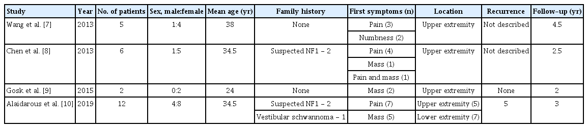
Characteristics of patients with segmental schwannomatosis reported in the previous literature
Regarding complications associated with the surgical excision of schwannomas, the incidence of postoperative neurological deficit is approximately 5% to 15%, with a low recurrence rate [4]. A sensory deficit, or at least a tingling sensation, after surgery may be more likely in cases with multiple schwannomas in the common and proper digital nerves. However, such neurological complications can be avoided by meticulous dissection and separation of the tumors from the nerve fibers under loupe magnification. Although recurrence was reported in five out of 12 cases of schwannomatosis in Alaidarous et al.’s most recent report [10], our patient did not present with recurrence in either the forearm or the hand.
In conclusion, we present a rare case of segmental schwannomatosis featuring multiple schwannomas in the common and proper digital nerves, 15 years after a huge solitary schwannoma was excised from the forearm. The final diagnosis was reached by careful surgical resection and pathological examination, and no complications were noted at the 1-year outpatient follow-up.
Notes
The authors have nothing to disclose.
Funding
None.