Surgical treatment of sagittal band injuries and classification according to operative findings
Article information

Abstract
Purpose
The indications for surgery in patients with acute closed sagittal band injuries are still undetermined. The purpose of this study was to classify the types of injuries based on intraoperative findings of patients who underwent surgery for sagittal injuries, and to present the treatment plans and surgical methods.
Methods
Twenty-five patients who had undergone surgical exploration for closed sagittal band injuries between January 2011 and December 2020 were included in the study, comprising 17 patients with acute injuries (within 3 weeks), four patients with chronic injuries, and four patients who underwent surgery because symptoms did not improve in response to conservative treatment. Patients with laceration, fracture, and rheumatoid arthritis were excluded.
Results
Sagittal band injuries were classified into two groups: superficial sagittal band (SSB) and proper sagittal band (PSB) injuries. SSB injuries were observed in 75.0% of spontaneous rupture cases and PSB injuries were observed in 66.7% of traumatic rupture cases. SSB injuries were observed in 83.3% of Rayan and Murray classification type II cases and PSB injuries were observed in 61.5% of type III cases (p=0.041). PSB injuries were present in all four patients who underwent surgery because conservative treatment failed.
Conclusion
We successfully corrected sagittal band injuries with extensor digitorum communis tendon instability through surgical treatment. Sagittal band injuries can be classified into two types depending on the anatomical injury pattern; SSB and PSB injuries. The surgical method and treatment plan can be chosen based on this classification.
Introduction
Among the structures constituting the extensor hood at the metacarpophalangeal (MP) joint level, the sagittal band plays the most important role in ensuring that the extensor digitorum communis (EDC) tendon is centered without being shifted to one side [1]. The sagittal band inserts into the extensor hood from the volar plate of the MP joint and the deep transverse ligament. In the groove of the metacarpal head, a thick deep layer and a thin superficial layer crossing the EDC tendon surround the tendon. The sagittal bands on the radial and ulnar sides are symmetrical to each other to prevent subluxation of the EDC tendon during flexion and extension of the MP joint.
Sagittal band rupture can occur under various conditions, such as compression trauma to the MP joint due to a direct blow or low-energy trauma such as finger flick [2]. It usually causes instability of the EDC tendon along with swelling and pain. Rayan and Murray [3] classified the severity of closed sagittal band injury into three types based on the degree of instability of the EDC tendon by subluxation and dislocation. Ishizuki [4] defined the difference between traumatic rupture and spontaneous rupture in closed sagittal band injury. Spontaneous rupture due to snapping and crossed fingers has been described previously. However, these classifications do not provide appropriate treatment guidelines. In addition, some cases did not fit the existing classification wherein surgery was performed for sagittal band injury at our clinic. Based on the intraoperative findings, we intend to define the injury type according to the anatomical injury pattern and suggest treatment plan.
Methods
Ethics statement: This study was performed in accordance with the Declaration of Helsinki. The need for patient consent was waived owing to the study’s retrospective nature, and the study design was approved by the Institutional Review Board of Gwangmyeong Sungae General Hospital (No. KIRB-2021-N-003).
1. Patients
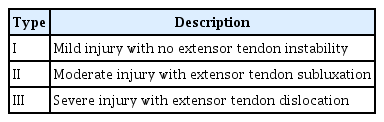
A retrospective study was conducted on 25 patients who were diagnosed with sagittal band injury and underwent surgery between January 2011 and December 2020 (Table 1), including 17 patients with acute injury within 3 weeks who decided to undergo early surgical treatment for immediate symptom improvement, four patients who underwent surgery due to no improvement of symptoms after conservative treatment, and four patients with chronic injury for more than 3 weeks at the time of admission. Fractures, lacerations, rheumatism, and other associated soft tissue diseases were excluded. The causes of injury were traumatic rupture and spontaneous rupture [4]. Among the injury mechanisms of the patients included in this study, those that occurred after finger flicking, and those that occurred after making kimchi or lifting a heavy box were classified as spontaneous rupture. On the other hand, injuries caused by hitting a wall with a fist or hitting the edge of an exercise device or desk edge were classified as traumatic rupture. For the degree of instability of the EDC tendon in the MP joint, we referred to the Rayan and Murray classification [3]. Type I was defined as mild injury with no extensor tendon instability; type II as moderate injury with extensor tendon subluxation; and type III as severe injury with dislocation of the extensor tendon.

Patient demographics, ultrasonography (USG), and intraoperative findings
This study was performed in accordance with the Declaration of Helsinki. The need for patient consent was waived owing to the study’s retrospective nature, and the study design was approved by the Institutional Review Board of Gwangmyeong Sungae General Hospital (No. KIRB-2021-N-003).
2. Intraoperative findings
Two types of injuries were observed based on the actual anatomical location of the injury through the surgical findings of patients with sagittal band injury. The first type was a torn superficial layer, which was classified as a superficial sagittal band (SSB) injury (Fig. 1). The second type included intact superficial and deep layers; however, damage was observed in an indistinguishable area due to the merging of the two layers on the lateral side. Thus, it was named the proper sagittal band (PSB) to distinguish it from the two layers and to classify it as PSB injury (Fig. 2).
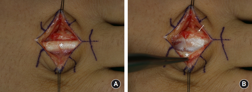
Superficial sagittal band injury. (A) Tear of the superficial layer of the radial sagittal band is noted intraoperatively. With the metacarpophalangeal joint in an extended state, the extensor digitorum communis (EDC) tendon (asterisk) remains in place. (B) The forceps are holding the EDC tendon (asterisk). The EDC tendon is detached from the deep layer. The deep layers beneath the EDC tendon and the proper sagittal band (arrow) on the radial side are intact.

Proper sagittal band (PSB) injury. (A) Complete rupture with gap formation on the ulnar side PSB (arrow) is noted intraoperatively. With the metacarpophalangeal joint flexed at approximately 15°, the gap widens and the extensor digitorum communis (EDC) tendon (asterisk) is displaced radially as a unit with the extensor hood. (B) The forceps are holding the thick PSB (arrow). The EDC tendon (asterisk) is completely covered by a superficial layer and a deep layer.
3. Surgical procedure and postoperative management
The surgeries were performed under regional anesthesia with brachial plexus block. The sagittal band was exposed by making an arc-shaped incision on the radial or ulnar side of the MP joint. The location and pattern of damage to the sagittal band were examined through the exposed area, and the degree of dislocation and subluxation of the EDC tendon was inspected while passively flexing the MP joint of the affected finger. Treatment was based on the direct method and differed according to the SSB and PSB injuries. The SSB injury was very thin compared to the thickness of the injured area, and the EDC tendon was intact in the deep layer under the MP joint extension. To immobilize the EDC tendon during MP joint flexion, the paratendon was sutured to the PSB by continuous method fixation using PDS 5-0 sutures (Ethicon, Inc., Somerville, NJ, USA) (Fig. 3A). The PSB injury was repaired by approximating both rupture margins and combining several figure-of-eight methods and continuous methods using PDS 4-0 or 5-0 sutures to prevent the gap from widening during MP joint flexion (Fig. 3B). If approximation was not possible due to severe disruption of the margin, or if tendon instability persisted even after direct repair, the tendon slip method was used to reconstruct it.
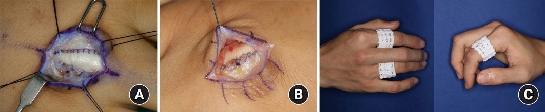
(A) The superficial sagittal band injury is slightly and loosely repaired using a paratendon PDS 5-0 suture (Ethicon, Inc., Somerville, NJ, USA) at the border of the extensor digitorum communis tendon. (B) In a case of proper sagittal band (PSB) injury, the ruptured PSB is substantially repaired to prevent gap widening through the multiple figure-of-eight method and the consecutive method using PDS 4-0 and PDS 5-0 sutures.
Immediately after surgery, passive flexion of the MP joint was performed up to 90° in all patients to confirm that the EDC tendon was not subluxed and stability was maintained. Postoperative management was maintained for 3 weeks after changing the immobilization splint to a dynamic splint (Fig. 3C) within 1 week after surgery, without distinguishing between SSB and PSB injuries. After confirming that EDC tendon instability did not recur, it was changed to buddy taping, and full range of motion was started.
The patients’ demographics, clinical findings such as EDC tendon instability, and intraoperative findings were retrospectively investigated while evaluating their medical records and photographs. The Fisher exact test was performed for categorical variables. In the interpretation of results, P-values of <0.05 indicated statistical significance. All statistical analyses were performed using IBM SPSS ver. 28.0 (IBM Corp., Armonk, NY, USA).
Results
The patients’ clinical characteristics are shown in Table 2. There were 21 male and four female patients, with a mean age of 30.7 years (range, 15–75 years). The middle finger was present in 24 cases and the index finger in one case. There were 16 cases of spontaneous rupture and nine cases of traumatic rupture. Instability of the EDC tendon was observed in all patients. Rayan and Murray classification types II and III were noted in 12 and 13 patients, respectively. The average period from symptom onset to surgery was 5.5 weeks (range, 1–52 weeks). Early surgery was performed in 17 cases with acute injury within 3 weeks (68.0%) and eight cases with chronic injuries (32.0%), including four cases in which conservative treatment failed. USG was performed preoperatively in 15 patients, and sagittal band injury was confirmed through EDC tendon instability, with an increase in the hypoechoic area (Fig. 4). Of the 25 cases, 22 were corrected through direct repair, and three were reconstructed using the tendon slip method. The average follow-up period after surgery was 6.6 weeks (range, 2–49 weeks), and in all 25 cases, no recurrence of tendon instability or impairment of range of motion occurred.
Patients’ clinical characteristics
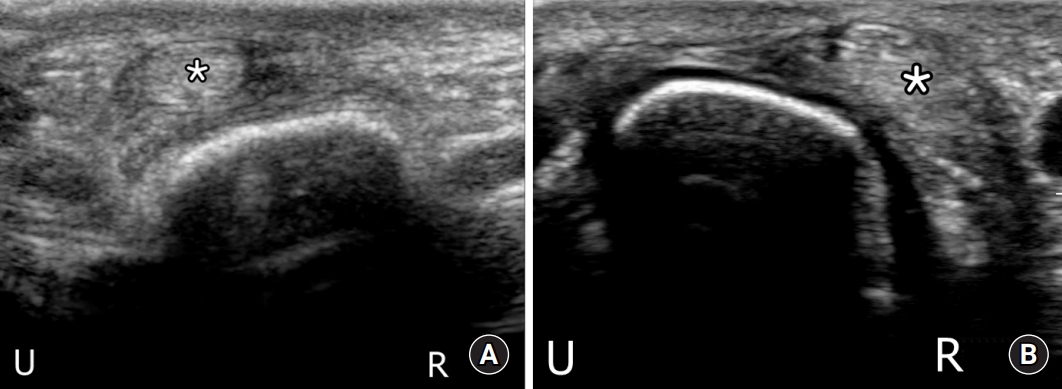
(A) Preoperative ultrasonography (USG) of a superficial sagittal band injury. Axial dynamic view of the third metacarpophalangeal (MP) joint during flexion shows hypoechoic thickening on the radial side of the extensor digitorum communis (EDC) tendon (asterisk). The EDC tendon is subluxed to the ulnar side. (B) Preoperative USG of a proper sagittal band (PSB) injury. Axial dynamic view of the third MP joint during flexion shows radial dislocation of the EDC tendon (asterisk). R, radial; U, ulnar.
All 25 sagittal band injuries included in this study were of two types; SSB and PSB injuries. Of the 25 cases, there were 15 cases of SSB injury and 10 cases of PSB injury. The correlation between our classification type and the existing classification was analyzed (Table 3). Of the 16 spontaneous rupture cases, 12 (75.0%) had SSB injuries and four (25.0%) had PSB injuries. Of the nine traumatic rupture cases, three (33.3%) were SSB injuries and six (66.7%) were PSB injuries. In the preoperative evaluation, according to the Rayan and Murray classification, of the 12 type II cases, 10 (83.3%) were SSB injuries, and two (16.7%) were PSB injuries. Of the 13 type III cases, five (38.5%) were SSB injuries and eight (61.5%) were PSB injuries. The more severe the instability to dislocate the tendon, the higher the probability of PSB injury, which was statistically significant (p=0.041).

Analysis of intraoperative and clinical findings
Depending on the location of the injury, the radial sagittal band was damaged in 19 cases and the ulnar sagittal band in four cases. In two cases, both sagittal bands were damaged. In both cases, partial SSB injury was found on the ulnar side, and complete SSB injury was observed on the radial side as the main injury, causing ulnar subluxation during the MP joint flexion. Damage was observed on the radial side in all 16 cases of spontaneous rupture. Although the radial side was intact, all four cases with sagittal band damage on the ulnar side were PSB injuries due to traumatic rupture. There were a total of eight chronic injuries 3 weeks after the onset of symptoms, including four patients with PSB injury who underwent surgery because symptoms persisted even after conservative treatment.
Discussion
Classification according to the actual injury location is more suitable in providing treatment guidelines beyond simple evaluation of the diagnosis and severity. Ishizuki [4] reported that spontaneous rupture was only involved in the superficial layer at the insertion site on the radial side of the tendon, whereas traumatic rupture involved both superficial and deep layers. In our study, the surgical findings of 10 cases revealed that rupture was observed on the lateral side slightly more than when the deep and superficial layers were separated. In the literature, this region is referred to as the radial sagittal band or the ulnar sagittal band as an extension of the superficial and deep layers. However, the two layers merge on the border of the EDC tendon and cannot be separated from each other. Thus, it was anatomically more appropriate to distinguish and define it from the superficial and deep layers. Therefore, this structure was named the PSB. Only the superficial layer was torn off in the other 15 cases.
All cases included in this study showed tendon instability. SSB injury was observed in 10 cases (83.3%), as a result of the surgical findings of 12 type II patients. The surgical findings of 13 type III patients showed eight cases of PSB injury (61.5%). If the instability was severe enough to dislocate the EDC tendon, there was a high probability of PSB injury. In spontaneous rupture, SSB injury was mainly observed in 75.0% of cases, and in traumatic rupture, PSB injury was observed in 66.7% of cases. Spontaneous or traumatic rupture may be associated with anatomical injury types, but are not statistically significant.
Differentiation between SSB and PSB injuries through preoperative examination is still insufficient. Among preoperative examinations, USG is useful for the diagnosis and evaluation of severity. In USG, the extensor hood, including the sagittal band, is best seen at the MP joint under 30° flexion, and in the axial view, the sagittal band appears as a thin hypoechoic region around the EDC tendon [1,5]. Pinpointing the exact location of damage under USG is still difficult. In evaluating EDC tendon instability, identifying severe symptoms, such as dislocation, with the naked eye is not as difficult as identifying mild symptoms. In particular, acute traumatic injury is more difficult to distinguish in the early stages of severe swelling. Through dynamic evaluation under USG, increased subluxation and dislocation of the tendon can be observed during finger flexion, making objective evaluation of EDC tendon instability possible [6]. When the mechanism of damage is traumatic injury, ulnar-side sagittal band injury, and instability large enough to dislocate the EDC tendon, PSB injury may be considered by confirming the discontinuity of the hypoechoic region lateral to the EDC tendon through USG.
Various surgical methods are known for the treatment of the sagittal band, and among them, the classic surgical method is to suture the tendon with the fiber of the damaged sagittal band. Hong et al. [7] successfully treated 26 type II and III patients with spontaneous ruptures through direct repair. Tendon slip or juncturae tendinum is mainly used when direct repair is not possible due to the tissue gap or chronicity of the damaged sagittal band [8,9]. After surgery, it is usually placed in an extension position and immobilized for 3 to 4 weeks, and in some cases, a dynamic splint is applied immediately after surgery [10]. In the surgical findings of this study, it was confirmed that the EDC tendon could not be supported at the site where the superficial layer was ruptured, and the tendon detached from the deep layer in SSB injury. For this reason, instability of the EDC tendon occurred during flexion of the MP joint. In PSB injury, a gap is formed as the PSB is completely ruptured on the lateral side of the EDC tendon. During MP joint flexion, the gap widens, and the EDC tendon is maintained as one unit with the extensor hood, and displacement occurs on the unaffected side. Based on these findings, a difference was made between the SSB and PSB injuries, and in the case of the PSB injury, the gap was repaired tightly to prevent it from widening.
In general, sagittal band injury is treated nonsurgically for acute injuries within 3 weeks, and surgery is performed when chronic injuries or conservative treatment do not improve. However, treatment guidelines are still insufficient, and for conservative treatment and surgery have been reported in various studies, especially in cases of acute injury [3,4,11-15]. In this study, PSB injury was observed in all four patients with acute injury who underwent surgery because symptoms persisted despite conservative treatment. We believe that early surgical treatment may be a good option for PSB injury. However, there is a limit that there are too few cases to present sufficient evidence, so additional study is needed in the future.
We classified the sagittal band injury as SSB or PSB based on the surgical findings according to the anatomical patterns of 25 patients, and although sufficient evidence is not yet supported, we would like to present a treatment plan (Fig. 5). For chronic injuries, including those that do not show improvement even after conservative treatment, surgery is still the treatment of choice. However, in the case of acute injury within 3 weeks, conservative treatment is started for SSB injury, but surgical treatment may be recommended for PSB injury. However, preoperative evaluation data to distinguish between SSB and PSB injuries are still incomplete. The relationship between the injury mechanism and the severity of EDC tendon instability has been confirmed to some extent, and if imaging techniques, such as USG and MRI, are developed along with these clinical findings and further research proceeds, SSB and PSB injuries can be distinguished from each other.

Treatment algorithm for sagittal band injuries. SSB, superficial sagittal band; PSB, proper sagittal band.
Conclusion
We successfully corrected the sagittal band injury with EDC tendon instability through surgical treatment. Sagittal band injury can be classified into two types depending on the anatomical injury pattern; SSB and PSB injuries. Surgical method and treatment plan can be chosen based on this.
Notes
The authors have nothing to disclose.
Funding
None.