전가쪽 대퇴부 역류 유경피판을 이용한 무릎주위 피부연조직결손의 재건: 증례 보고
Reconstruction of Soft-Tissue Defect Around the Knee Using a Pedicled Reverse-Flow Anterolateral Thigh Flap: A Case Report
Article information

Trans Abstract
Coverage of traumatic soft-tissue defects around the knee is a challenging problem for reconstructive surgeons though many reconstructive options are available. We planned to use a pedicled reverse-flow anterolateral thigh (ALT) flap using the distal branch of the descending branch of the lateral circumflex femoral artery (LCFA) for pedicle length extension in a patient with the ALT perforator branch originating from the proximal portion of the descending branch of LCFA. We present the successful use of a pedicled reverse-flow ALT flap to cover a soft tissue defect around the knee.
Introduction
Soft tissue coverage around the knee after trauma remains a challenge for plastic and reconstructive surgeons. The anterolateral thigh (ALT) flap was first described in 1984 by Song et al. [1] and has become the most commonly used perforator flap due to its versatility, ease of use, and minimal donor site morbidity [2,3]. Perfusion of the ALT flap is based on the musculocutaneous or septocutaneous perforators of the descending branch of the lateral circumflex femoral artery (LCFA) [4], which connects distally with the lateral superior genicular artery or the profunda femoral artery [4]. The pedicled reverse-flow ALT flap was first described by Zhang [5], who used a retrograde vascular pedicle to prevent active bleeding from the distal end of the descending branch of the LCFA. In the described case, we planned a pedicled reverse-flow ALT flap for pedicle length extension with the distal descending branch of the LCFA to address possible insufficient pedicle length because the perforator emerged from a proximal portion of the descending branch.
Case Report
A 69-year-old female had a left tibia plateau open fracture with large degloving injury resulting from a traffic accident. After several surgical debridements, the resultant defect measured 20.0 cm×15.0 cm with significant patella tendon exposure (Fig. 1). Prior to surgery, a perforator pedicled propeller flap using a musculocutaneous or septocutaneous perforator branched from the distal portion of the thigh was planned. Doppler mapping of perforators was performed before flap elevation. The method used was similar to the conventional method used to harvest an ALT flap, that is, with the identified perforator in the center, a 12.0 cm× 4.0 cm skin paddle was designed to cover the exposed patella tendon of the anterolateral knee. A search for cutaneous perforators was performed in the suprafascial and subfascial plane, but we failed to identify a perforator branched from distal portion of thigh intraoperatively. The only musculocutaneous perforator found branched at the proximal descending branch of the LCFA 12.0 cm above the lateral edge of the patella. So, the preoperative plan was changed to a pedicled reverse-flow ALT flap. The identified perforator was dissected in a retrograde fashion, and the proximal pedicle was clamped using a vascular clamp and the flap pedicle was isolated distally along the descending branch until a pivot point was reached 7 cm above the knee joint that allowed sufficient pedicle length without tension. Intramuscular dissection of the perforator with skeletonization of the descending branch helped increase the degree of flap advancement, and the pedicled reverse-flow ALT flap was elevated and transposed to cover the exposed patella tendon. The pedicle length was 10 cm (Fig. 2A, 2B). The donor site was closed primarily and the other skin defect area around the knee without tendon exposure was covered with a split thickness skin graft from the right thigh. The wound healed completely without any complication and remarkable limitation of range of movement after 3 months (Fig. 3).

A soft tissue (20.0 cm×15.0 cm) defects above the knee following several debridement, with significant patella tendon exposure on anterolateral side of the knee.
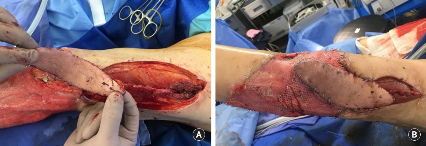
(A) A pedicled reverse-flow anterolateral thigh flap (12.0 cm×4.0 cm sized) was harvested using musculocutaneous perforator of descending branch of the lateral circumflex femoral artery (LCFA). Descending branch of the LCFA and the perforator to the flap was dissected intramuscularly. (B) The pivot point was determined at about 8 cm above the knee joint and the pedicled reverse-flow anterolateral thigh flap was advanced to cover the patella tendon exposure area.
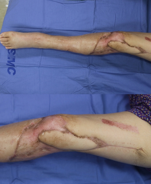
Photograph of the flap at 3 months of follow-up. Satisfactory healing and contour achieved.
Written informed consents were obtained.
Discussion
Coverage of traumatic soft-tissue defects around the knee remains a challenging problem for reconstructive surgeons because of a lack of an adequate recipient vessel in anterior, lateral knee area and the need for a thin, pliable knee joint coverage. Various options have been utilized to provide soft tissue reconstruction for these defects. The use of a free flap is not always preferable in this region, due to high donor site morbidity, long surgical time, and difficulties associated with selecting an appropriate recipient vessel positioned deeply around the knee. Pedicled muscle flaps have been the workhorse for coverage of defects around the knee for decades. The gastrocnemius muscle flap has proved to be the most reliable, safest, and easiest surgical option [6]. However, perforator flaps have recently revolutionized soft tissue reconstruction around the knee, due to low donor morbidity and available pedicle length. Gravvanis et al. [7] demonstrated the use of a pedicled reverse-flow ALT flap for knee reconstruction and considered it super to the gastrocnemius muscle flap, because of its greater size, shape, and flexibility, better color and texture match, and less bulkiness. Furthermore, pedicled reverse-flow ALT flap have arcs of rotation long enough to reach suprapatellar defects and are capable of resurfacing the whole knee. However, it is technically difficult to dissect ALT perforator flaps, especially when they are small, because perforating arteries exhibit wide anatomic variations. In 1998, Kimata et al. [8] classified the perforators of ALT flaps into 8 types according to the branching pattern of the perforator vessel.
The pedicled reverse-flow ALT flap is based on perforators of the descending branch of LCFA. Zhang [5] introduced the concept of a pedicled reverse-flow ALT flap in 1990. However, despite its several advantages and low donor site morbidity, the pedicled reverse-flow ALT flap has not been widely adopted, possibly because of wide anatomic variations of the vascular pedicle and a high risk of venous congestion [9]. In addition, reverse-flow based flaps are prone to congestion in peripheral portions of flaps, and this is followed by partial necrosis, because of insufficient venous drainage caused by the resistance of venous valves. In a recent study, a modified method was described that reduces venous congestion, wherein the venae comitantes is anastomosed to the proximal stump in an anterograde manner [7,10]. Wong et al. [9] reported sixteen cases of pedicled reverse-flow ALT flaps. Of 16 cases, venous congestion was noted in 8 cases, 3 of which were salvaged with venous supercharging with the long saphenous vein. Two that were not venous supercharged underwent partial flap necrosis and all other flaps healed without complications. In our case, there was no sign of severe venous congestion, and thus, no additional venous augmentation was performed.
Originally, we planned a perforator pedicled propeller flap to cover the soft tissue defect around the left knee based on preoperative doppler mapping findings, but intraoperatively, we were unable to find the perforator near the midpoint of the thigh. We only found a musculocutaneous perforator from a proximal region of the descending branch of the LCFA, which prevented use of a propeller flap. Accordingly, the reconstruction plan was changed to a pedicled reverse-flow ALT flap with pedicle length extension to enable the elevated flap to reach the knee tendon extension area. To achieve this, the proximal branch of the LCFA was clamped and its distal branch was dissected to increase flap mobility sufficiently to reach the exposed knee tendon area without excessive tension.
Due to anatomic variations of ALT flap perforators, surgeons might encounter unplanned situations during surgery. If a planned flap cannot be applied, a decision must be made whether to adopt an alternative reconstructive method or some other method in same operative field. Notably, ALT flap pedicle lengthening might be required in same operative field and may aid reconstruction, and recent studies have described ALT flap pedicle length extension in operative fields [10]. The pedicled reverse-flow ALT flap is a good option that enables relatively straightforward pedicle extension within the same operative field when securing an adequate pedicle length is difficult because of a proximal location of the perforator in the descending branch.
As described in the present case, successful soft tissue reconstruction around the knee is possible using a pedicled reverse-flow ALT flap, especially when the perforator in the descending branch of the LCFA emerges from proximal thigh.
Notes
The authors have nothing to disclose.