Reconstruction of a high-energy penetrating injury from the abdomen to the sacral area using a latissimus dorsi free flap with monofilament polypropylene mesh and pedicled flap rotation: a case report
Article information

Abstract
A 50-year-old man was transferred to a level I trauma center for penetrating injury. Industrial metal had penetrated his trunk, and he was injured in internal organs. The injured internal organs were treated by the trauma surgery team. The peritoneum was reconstructed with artificial dermal matrix graft. The wound was managed with negative-pressure wound therapy, and several debridement procedures were performed. The full-thickness abdominal defect was covered with monofilament polypropylene mesh(Parietene mesh, 30×30 cm). A latissimus dorsi flap was elevated with a musculocutaneous flap measured 50×30 cm, and 6-cm thoracodorsal artery pedicle. Microvascular anastomosis was performed using the thoracodorsal and left femoral arteries. Two weeks later, we performed local flap rotation based on gluteal artery perforator in the sacral area. Polypropylene mesh was successfully inserted without complications. Combining a latissimus dorsi free flap on a polypropylene mesh can be an effective method for reconstructing large penetrating wounds on the trunk.
Introduction
Trauma is a major component of occupational accidents, leading to death and disability, and a critical contributor to decreased productivity. Reconstruction through free flap surgery for soft tissue defects caused by trauma is challenging for plastic surgeons in the field of trauma injuries. Penetrating injuries are rare but fatal and are often described as penetrating injuries with sharp materials [1,2]. Penetration injury can cause bowel injury accompanied by abdominal wall defects [3]. The methods used to perform abdominal wall reconstruction depend on the location, size, and thickness of the defect. The reconstruction of complex defects in the abdominal wall requires careful planning. Full-thickness, but limited defects are treated using various pedicled myocutaneous flaps, involving the tensor fasciae latae myocutaneous, rectus femoris muscle, myocutaneous, anterolateral thigh fasciocutaneous, sartorius muscle or myocutaneous flaps, and latissimus dorsi (LD) myocutaneous flaps.
Reconstructing large abdominal wall defects caused by malignant neoplasms has been previously reported [4]. However, life-threatening dorsally to ventrally penetrating injuries are very rare. This study presents our experience with the coverage of a large complex full-thickness abdominal wall defect due to traumatic penetrating injury with a free flap and mesh.
Case report
All procedures performed in this study involving human participants were in accordance with the ethical standards of the Ajou University Hospital and of the Declaration of Helsinki, as revised in 2013. Written informed consent was obtained from the patient for the publication of this case report and accompanying images.
A 50-year-old man underwent penetrating trauma by casting a shearing machine that extended from the abdominal area to sacrum. The trauma involved the colon, small bowel, abdominal wall, pelvic bone, and ureter (Fig. 1).
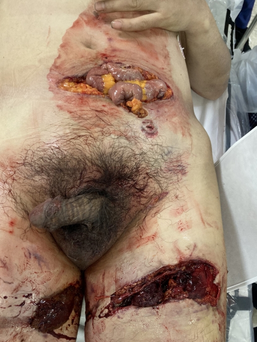
Initial status of the patient upon arrival at the trauma bay after a penetrating injury by a compressing machine.
Immediately after he was transferred to the level 1 trauma center, the trauma team, urologist, and general surgery team performed combined surgery, involving general, urological, and orthopedic surgery. The urologist performed end-to-end anastomosis of the left ureter and cystostomy, and the general surgery team performed the segmental resection of the damaged ileal and sigmoid colon, ileostomy, and wound debridement.
Subsequently, debridement and peritoneal irrigation of the dirty wounds were performed every 2 to 3 days by the trauma team (Fig. 2). Two weeks after trauma, the general surgery team implanted a 30×10-cm allogenic acellular dermal matrix into the abdominal wall (Fig. 3). A wide range of injuries and accompanying bowel injuries and infections may have resulted in sepsis. It took time for the wound to clear and the patient’s vital signs to recover, and colostomy was required to resolve the injured bowel and infection, warranting several days of follow-up. Reconstruction of the abdominal wall by the plastic surgery team was performed 46 days after trauma. Acellular dermal matrix and negative-pressure wound therapy can be helpful materials that can temporarily cover the time required for these treatments. The first defect of the abdominal wall was covered with a monofilament polypropylene mesh (Parietene mesh, 30×30 cm; Medtronic, Dublin, Ireland) to prevent ventral hernia. The LD flap was elevated, making a 20×10-cm fasciocutaneous flap on a 50×30-cm muscle flap, with a 6-cm thoracodorsal artery pedicle. A microvascular anastomosis was made in end-to-side fashion from one artery and one vein of the pedicle to the left femoral artery and vein (Fig. 4).

Wounds after 2 weeks of multiple wound perfusion and debridement. (A) Abdominal side. (B) Sacral side.
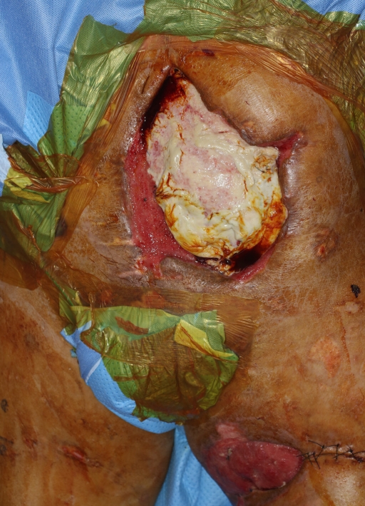
Abdominal wound applied with acellular dermal matrix for maintenance of the intestinal injury recovery and temporary intestinal coverage.
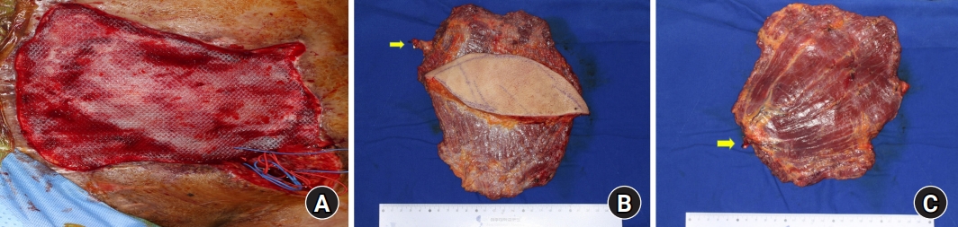
Abdominal wound repaired with mesh and latissimus dorsi musculocutaneous flap with monofilament polypropylene mesh coverage. (A) Wounds with monofilament polypropylene mesh applied. (B, C) Elevated latissimus dorsi musculocutaneous flap. Yellow arrows indicate the thoracodorsal artery.
The sacral wound was treated according to grade IV pressure injury because there was no long-term exposure of the internal organs. However, it was necessary to maintain drainage for a long period of time because of the generation of collection fluid due to wound penetration.
Numerous debridements and vacuum-assisted dressings were performed for the sacral area defect. After granulation tissue formation 60 days after the onset of trauma, the defect was covered by the advancement of the adjacent muscle flap, and the gluteal artery perforator-based local flap was rotated to cover the raw surface by the plastic surgery team (Fig. 5). After 6 weeks of sacral reconstruction, the patient recovered with adequate flap healing. The sacral area had no complications, and his sitting and lying down postures were correct.

Image at 2 months postoperatively. (A) Abdominal side. (B) Sacral side.
Discussion
Studies on abdominal wall reconstruction with artificial mesh materials and free tissue transfer in cases of traumatic abdominal wall disruption are limited. Ventral to dorsal penetrating injuries are rare, but they are fatal injuries due to the contusion of surrounding tissues and open windows of tissues. Damage is affected by the location, penetrating material, and penetrating energy. Particularly, industrial penetrating equipment causes destructive damage to surrounding tissues owing to high-energy damage. Abdominal high-energy penetrating injury is fatal and requires a multidisciplinary approach as it causes internal multiple organ damage, soft tissue defects, and fractures [5].
Various methods, including rectus muscle flap, anterolateral thigh flap, and component release, have been described for abdominal wall reconstruction [5]. Moreover, depending on the location of injury, a pedicled flap can be integrated using a perforator of the superior epigastric artery, inferior epigastric artery, or intercostal artery. Reconstruction using a musculocutaneous flap or muscle flap is often performed for complex high-energy injuries in the abdomen [4]. LD musculocutaneous flaps are often the only option, especially when the defect size is very large. When using the free flap, in addition to the arteries described above, the superficial femoral artery, with or without vein grafts, may be used [3-5].
Cases of penetrating abdominal injury are often polytraumatic. As it is accompanied by pelvic fracture or internal organ injury, delayed reconstruction is inevitable. If the wound is too large to be reconstructed or repaired, secondary reconstruction, including skin graft or flap, can be performed after a period of open dressing to allow granulation to grow over the intestinal loop. Negative-pressure wound therapy can be safely used for wound maintenance in acute vital recovery from concomitant injuries, including bowel exposure or pelvic fractures [6].
Due to the defect of the deep fascia in addition to extensive damage, the risk of hernia increases, even if the deep fascia is reconstructed with conventional free tissue transfer. Therefore, to reduce the risk of such a complication, we attempted to prevent secondary sequelae through mesh reconstruction. Coverage with an LD free flap with a polypropylene mesh is a possible option for extensive and full-thickness abdominal wall defects. Musculocutaneous flaps have an advantage in parietal repair owing to their characteristic aponeurotic components. In this case, the wound was not covered using any other free tissue transfer methods.
In the reconstruction of the complex abdominal wall including the abdominal wall in this case, there are few mentions of abdominal reconstruction using mesh after wide resection in the malignancy case [7]. However, in case of extensive damage due to high-energy penetrating phase damage, there are few descriptions due to high fatality. As in this case, ventral to dorsal penetrating injury required ileostomy, limiting the use of the pedicled artery in abdominal reconstruction, and the available vessels were limited due to a wide range of injuries.
In cases similar to the one presented herein, the use of a new prosthesis and polypropylene mesh may be a good option for patients with penetrating abdominal injuries, allowing a low complication rate.
Notes
The authors have nothing to disclose.
Funding
None.
Acknowledgements
We thank our patient for providing consent for the publication of this article.