Introduction
The radial forearm free flap has been commonly used for intraoral reconstruction because of its constant vascular anatomy and pliable skin texture. However, a few cases of anomalous branches of the radial artery have been reported. We report an anomalous superficial radial artery (SRA), detected during the elevation of a radial forearm free flap. A novel technique was used to preserve the circulation of the entire flap.
Case report
A 46-year-old 12-pack-year hypertensive man was diagnosed with squamous cell carcinoma of stage T3N0M0 on the left margin of the tongue, extending 3 cm deep as observed on magnetic resonance imaging. Left side hemiglossectomy, including the base of the tongue, floor of the mouth, and partial genioglossus muscle, was performed via lingual release and neck dissection. The pathologic findings revealed a 1.9×1.7 cm squamous cell carcinoma with an 8 mm depth of invasion. There was no lymphovascular invasion, but perineural invasion was noted. A radial forearm free flap was planned to directly cover the defect. The patient was right-handed, and a preoperative Allen’s test of the left hand showed adequate blood flow from both the radial and ulnar systems.
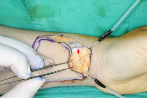
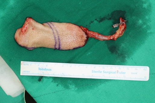
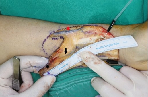
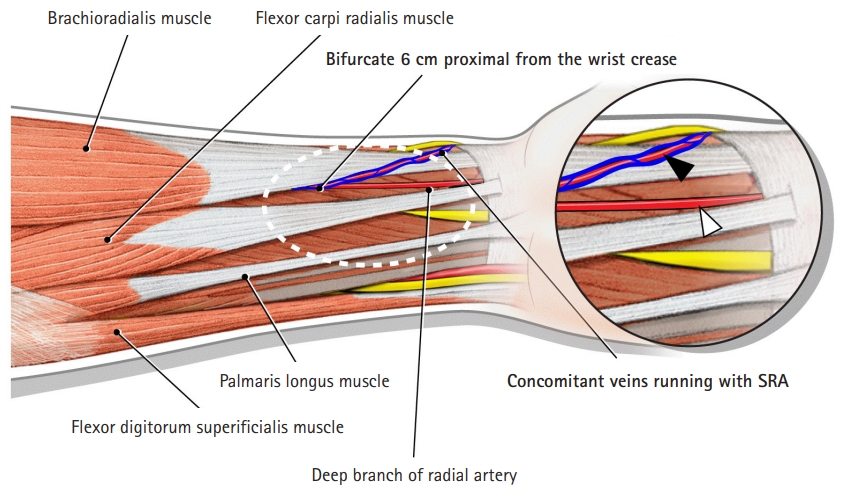
The flap was designed on the volar side of the patient’s hand. The flap measured 6×6 cm. The distal end of the flap, which became the tip of the tongue, was located on the radial artery at the level of the wrist crease. Exsanguination was performed using an Sterile Esmark Bandage (Medline Industries, Inc., Mundelein, IL., USA) and pneumatic tourniquet. First, the palmaris longus tendon and flexor carpi radialis tendon were identified at the level of the wrist crease. Then, the radial artery was clipped. The diameter of the radial artery measured 1.5 mm, which was smaller than the average diameter. Another dominant branch of the radial artery was considered. Upon proceeding with the flap elevation, an artery that ran with two veins was located superficial to the brachioradialis and ulnar side of the cephalic vein (Fig. 1). This aberrant artery was identified as the SRA. The SRA had a diameter of 2.0 mm, and its flow was more dominant than that of the clipped deep branch of the radial artery. The flap design required modification to ensure the circulation of the entire flap. The distal end of the SRA became the tip of the neotongue, and the flap design was extended 3 cm proximally to secure a sufficient flap size (Fig. 2). The superficial and deep branches of the radial artery bifurcated 6 cm proximal to the wrist-crease level. Consequently, both the superficial and deep branches of the radial artery were included in the flap (Fig. 3). A schematic illustration shows the course of deep and superficial branches of radial artery (Fig. 4). Flap transfer and insetting were accomplished without complications (Fig. 5). The patient recovered with adequate healing of the flap and donor areas. No hand function deficits or subjective complaints were noted.
Written informed consent was obtained for publication of this case report and accompanying images.
Discussion
Radial artery anomalies have been previously reported [1]. At the level of the forearm, a large branch arising from the radial artery at the proximal region 5 to 7 cm from the wrist is called the SRA [2,3]. It bifurcates from the deep branches at various locations. Its reported locations include the antecubital fossa [4], 10 cm distal to the antecubital fossa [5], middle of the forearm [6], distal 1/3 of the forearm [7], distal 1/4 of the forearm [1], and 4 cm from the wrist crease [8]. SRAs have been observed in 0.6% to 1.5% of cases [2]. It runs down the radial side of the forearm subcutaneously over the brachioradialis tendon, superficial to the anatomical snuff box, as it proceeds continuously through the dorsum of the hand [2,3]. During intravenous access, SRAs have been confused with the dorsal vein of the hand [9].
Radial artery anomalies can be identified preoperatively via radiographs, such as an angiogram or angiocomputed tomography, but they are not routinely performed. Physical examination, including the Allen’s test and pulse palpation, is the sole clinical method for predicting an aberrant artery. However, the preoperative physical examination does not confirm the bifurcation level, blood flow maintenance, or predominance of the aberrant artery. Therefore, large branches should not be divided during dissection until adequate exposure of the arterial system is achieved. The identification of anomalies facilitates harvest modifications to achieve successful anastomosis and flap perfusion. In cases wherein the SRA bifurcates at the proximal level, the flap is elevated, based on the aberrant artery, and another radial artery branch is preserved [10]. In this case, however, both radial artery branches were sacrificed to ensure flap circulation. In conclusion, despite the low incidence, surgeons should be aware of SRA or other aberrant branches of the radial artery, which may be located in various locations during radial artery-related flap elevation.