Introduction
Degloving injuries happen in various accidents, typically by roller machines [1]. A pull-out force with low velocity and moderate to high pressure destroys the loose connection between the superficial skin and deep tissues and separates the skin from underlying tendons, neurovascular and musculoskeletal units [1-4]. The goal of treatment is to preserve as many digits with functional length as possible. To achieve this goal, early coverage with good quality skin and early mobilization are imperative. In order to replace the large surface of the degloved hand, large flaps are usually needed. Despite various attempts, flaps from different donor sites have thicker skin than that of digits, and also cannot imitate the glabrous skin of the hand. Hence, functional reconstruction of degloved hand is challenging.
Obviously, replantation of the amputated stump is the best option. According to the classification of Urbaniak et al. [5], replantation is most effective in “class III” complete degloving amputations of fingers. However, in severely contused skin flaps direct microanastomosis may not be possible due to irreversible damage on vessel intima [1,3,5]. Other rescue options are defatted full-thickness skin graft (FTSG) or meshed split-thickness skin graft (STSG) over the avulsed surface, but these may not be possible in every case. This is because the crushed wound bed often presents compromised circulatory conditions, which hinder graft take and reperfusion of digits. Therefore, often wide necrotic change and amputations on digits are inevitable [1,2,5].
We present an experience in which a large volume of the amputated part was saved by composite graft, without any defatting or meshing procedure, in a totally degloved hand.
Case report
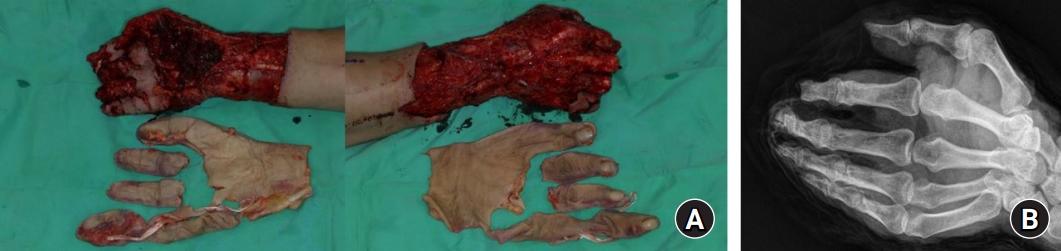
A 44-year-old man, injured by a rolling machine, came to the emergency room with a totally degloved right hand from wrist to fingertips. Nearly the entire skin from wrist to fingertips of thumb to ring fingers was detached, except for the small finger and a small portion of skin on the palmar side of the 3rd and 4th metacarpophalangeal (MCP) joint. The plane of separation was at the suprafascial level, leaving most of the muscles and neurovascular bundles intact. However, severe crushing was seen on almost the entire wound bed, especially on thenar region and around MCP joints of digits. In plain X-ray, bony amputation was shown on the index finger at the distal interphalangeal (DIP) joint level, and on the distal phalanx shaft level of the thumb, middle, and ring fingers (Fig. 1).
Avulsed skin was severely contused and torn apart into four pieces. Before exploration, copious irrigation with normal saline was done on the avulsed skin, but no other disinfectant solutions were used. Under a microscope, the vessels were not all located on the same plane but most vessels were left on the right hand. Most arteries and veins, including palmar digital arteries, were ruptured or suffered a severe intimal contusion. Replantation was not possible, therefore we carried out direct composite grafting of the untailored avulsed skin on the exposed hand and fingers. Since the chance of survival of the avulsed skin was obscure, our initial purpose was to prevent deep tissue dryness, which brings necrosis to bone and tendons. Hence, defatting or thinning by mesh was not needed. After composite grafting, mild compressive occlusive dressing was applied to keep the tissue under moist conditions.
Postoperatively, instead of negative pressure wound therapy, a daily change of dressing was done. Prostaglandin E-1 (alprostadil; Welfide Corp., Osaka, Japan) 10 μg daily was given intravenously for the first 7 days. Cefazedone 1 g and netilmicin 150 mg were given intravenously for 2 weeks.
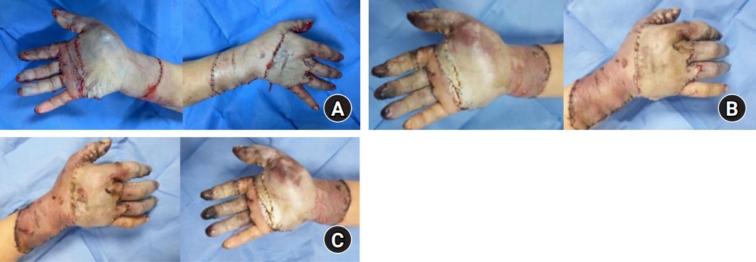
Immediately after surgery, the skin flap was pale without any sign of circulation, and blanching was not found. However, on postoperative day (POD) 4, the skin showed a bright red hue around the dorsum of the hand and proximal phalangeal level of digits. Until the first week, the red hue maintained its brightness without turning into dim and dark color. The escharic changes represented by dryness with marginal discharge were not shown. At POD 7, the area showing red hue extended to the palmar side of hand and vague blanching was seen on the adapted skin (Fig. 2). Until POD 10, skin maintained softness and those regions that showed red hue gradually turned into an evident pinkish color. We could not test the skin turgor in the early PODs since slight pinching of the grafted tissue could cause detachment and interfere with graft take.
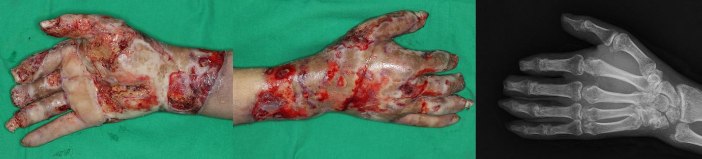
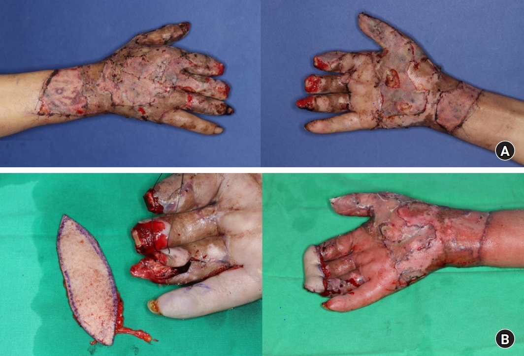
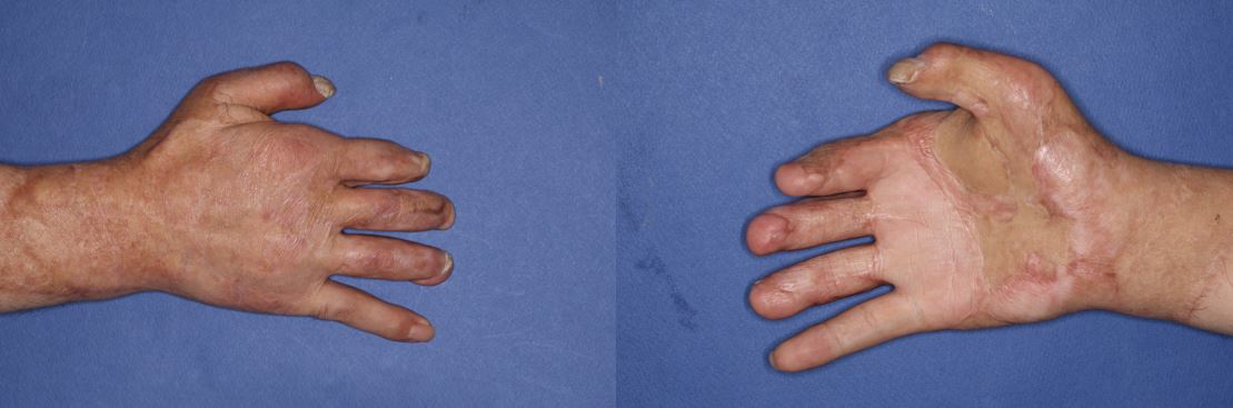
Since the extent of skin survival was obscure, we waited until the 5th week for debridement. At the 5th week, 65% of the surface area of adapted stump fully survived, with losses mostly by de-epithelization on palm and dorsum of the hand, and on the volar radial side of an index finger. Demarcation with volume changes on the distal phalanx of thumb, index, middle, and ring finger started and gradually progressed from POD 10. At the 5th week, minimal open amputation was done at the fingertip of the thumb, and distal to DIP joints of the index, middle, and ring fingers (Fig. 3). Skin defect on the hand dorsum and palmar side was covered by STSG from the thigh. Then, exposed DIP joint surfaces of the index, middle, and ring fingers were covered by a single anteromedial thigh (AMT) bridged free flap, of which painful fingertips were later revised by delayed the second toe pulp free flap (Figs. 4, 5).
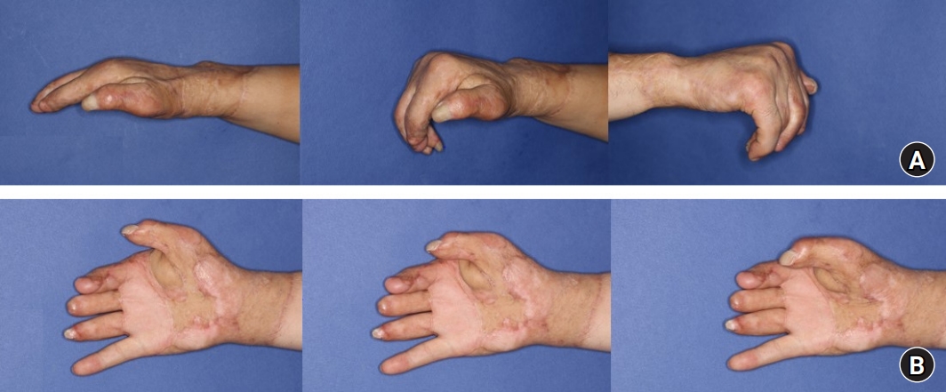
To prevent joint stiffness, early active and passive range of motion on MCP and proximal interphalangeal (PIP) joints was encouraged as soon as pain and swelling subsided. A short arm splint was self-removed during the daytime while undergoing exercise. After discharge, more aggressive passive exercise was done by a physical therapist three times a week in the outpatient clinic. Finally, the index, middle, ring, and small finger reached nearly 90° at active flexion and 10° at the extension on MCP joints, and 40° at flexion and 20° at the extension on PIP joints. For the thumb, MCP joint showed 70° at flexion and 40° at extension. Besides, 40° of radial abduction and nearly 60% of normal opposition were recovered. Although there were limitations in active range of motion and intrinsic motor functions, the skin maintained pliability without any signs of significant contracture (Fig. 6).
Written informed consent for the publication of clinical images were obtained from the patient.
Discussion
Lo et al. [4] classified the upper extremity degloving into two; expendable or non-expendable areas. They asserted injuries on functionally important, non-expendable areas like fingers need aggressive replantation. Urbaniak et al. [5] also asserted that replantation is most effective in “class III” complete degloving amputations of fingers. In our case, replantation seemed to be the best option, since nearly all fingers and hand were denuded. However, we could not find any repairable vessels on the avulsed skin. Unlike conveyor belts, roller machines have uneven grasping force, and thereby the force is applied to the axilla. This usually causes brachial plexus injury, but once degloving on hand occurs, roller machines cause severe contusion and crushing on vessel intima [3,5].
As an alternative, free flap or pedicled flaps may be considered. However, they are hardly attractive options due to donor morbidity (scarring and stiffness), longer operation time, and limitations by patient’s comorbidity. Moreover, the thick skin of these donors cannot replace the glabrous skin of the hand [2-4].
This leaves us with the last resort, defatted FTSG or meshed STSG of the avulsed skin. Coverage with defatted skin is a common measure, since it is known that defatting promotes graft take and decreases the chance of infection. Truly these are simple, affordable, and effective options for partial degloving injuries with the proximal or distal end still attached to the bed. However, on totally degloved hands, skin grafts have high necrosis rates and commonly cause rapid contracture by a lack of sufficient volume [5]. Therefore, it is hard to gain functionally viable digits through thinned skin grafts [6]. In our case, although replantation was not possible, we directly readapted the untailored avulsed skin, without defatting or meshing. We could not predict the extent of survival, and planned for free flap afterward.
Our attempt of composite grafting the untailored avulsed skin on a totally degloved hand, met with survival in a large volume of the amputated stump, while preserving the functional length of digits. Besides, the skin maintained pliability and showed less contracture than it would be presented by skin grafts. We have done minimum amputations on fingertips of the index, middle, and ring fingers, enabling coverage with a single AMT-bridged flap and delayed partial second toe pulp flaps. Despite a single case, surgeons facing disastrous total degloving amputation on hand may also consider direct composite grafting, rather than defatted skin grafts in case when replantation is impossible.